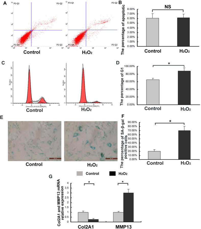
Figure 4. Increased levels of degenerative human CEP cell senescence resulting from oxidative stress induced by 150 μM H2O2.
(A) Sublethal oxidative stress induced by H2O2, and the early-and late-apoptosis of CEP cells were detected with Flow cytometry. (B) The addition of H2O2 did not increase CEP cell apoptosis. (C) Sublethal oxidative stress induced by H2O2, and the cells in different cycles including G1, G2 and S were counted and represented as a percentage of the total cell count with Flow cytometry. (D) The addition of H2O2 increased the population of G1 phase cells. (E) Sublethal oxidative stress induced by H2O2, and CEP cells were detected with SA-β-Gal staining. (F) The addition of H2O2 increased the percentage of SA-β-Gal-positive cells (blue) and the staining intensity. (G) The addition of H2O2 decreased Col2A1 expression and increased MMP13 mRNA expression. Three independent experiments were performed on the degenerative human CEP cells from three patients, and the data was denoted in terms of mean ± SD. *P < 0.05. NS: P > 0.05.

