Figure 2.

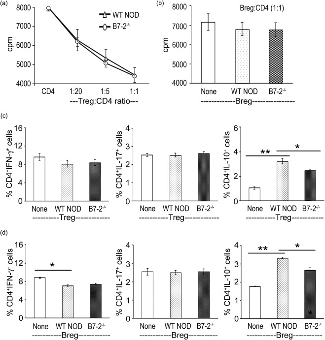
Effect of CD4+ regulatory T cells (Tregs) and regulatory B cells (Bregs) on T cell function in vitro. (a) Wild‐type (WT) and B7‐2–/– Tregs suppressed T cell proliferation with similar efficacy in vitro. CD4+CD25+ Tregs from 3‐month‐old female mice were co‐cultured with CD4+CD25– T cells from spontaneous autoimmune polyneuropathy (SAP) mice (8 months) in the presence of 20 μg/ml P0 (180–189) and irradiated antigen‐presenting cells (APCs) (50 000) for 3 days. Proliferation was measured by [3H]‐thymidine incorporation (n = 3). (b) WT and B7‐2–/– Bregs (CD19+CD1dhi CD5+) did not suppress the proliferation of CD4+CD25– T cells at a 1 : 1 ratio (n = 3). (c) WT and B7‐2–/– Tregs had no effect on CD4+interferon (IFN)‐γ or CD4+interleukin (IL)−17+ cells, but increased the percentage of CD4+IL‐10+ T cells in co‐cultures. Experimental conditions were the same as in (a), except that Tregs were obtained from WT or B7‐2–/– NOD mice killed at 10 days post‐immunization with P0 (180–199). Treg : CD4 ratio was 1 : 1. *P < 0·02; **P < 0·0005 (n = 3). (d) WT and B7‐2–/– Bregs induced a decrease in CD4+IFN‐γ+ T cells and increase in CD4+IL‐10+ T cells in co‐cultures. Bregs sorted from immunized animals were co‐cultured with CD4+CD25– T cells at a : 1 ratio for 3 days in the presence of P0 (180–199), lipopolysaccharide (LPS) (100 ng/ml) and irradiated antigen‐presenting cells (APCs) (50 000). *P < 0·002; **P < 0·0001 (n = 3).
