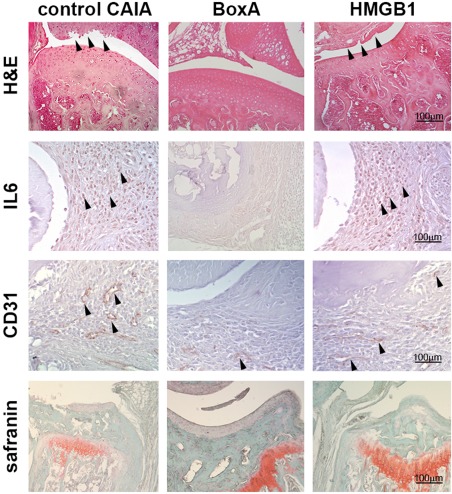
Figure 2.

Representative micrographs illustrating the haematoxylin and eosin staining, the immunohistochemical staining of interleukin (IL)‐6, CD31, a specific marker of endothelial cells and the safranin staining in control arthritic, BoxA‐treated and high‐mobility group box 1 (HMGB1)‐treated mice obtained on day 21 of arthritis. For immunohistochemical staining, positive staining appears in brown. Magnification ×200; n = 10 sections per joints; n = 5 mice per group.
