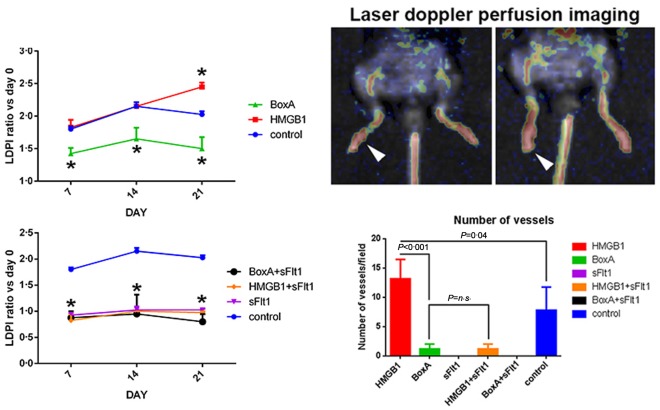
Figure 4.

Foot blood flow monitored in vivo by laser Doppler perfusion imaging (LDPI) in untreated control arthritic, BoxA‐treated and high‐mobility group box 1 (HMGB1)‐treated mice. Representative evaluation of an HMGB1‐treated mice 7 (left) and 21 days (right) after arthritis induction. In colour‐coded images, red indicates normal perfusion while yellow indicates reduced perfusion and blue indicates a marked reduction in blood flow in the hindlimb. The results are expressed as the ratio between the perfusion of the sum of the four limbs with that measured before the induction of arthritis. The blood flow of the arthritic joint is expressed as the ratio between perfusion of day 0 versus days 7, 14 and 21. *P < 0·05 versus control collagen antibody‐induced arthritis (CAIA) mice. The number of vessels per field, obtained by evaluation of the CD31‐positive cells, was increased significantly in HMGB1‐treated mice with respect to control CAIA mice; n = 5 mice per group.
