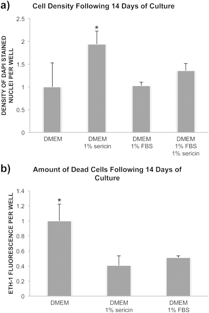
Figure 6. Effect of sericin on cell survival.

To assess the effect of sericin on cell survival confluent layers of human retinal pigment epithelial cells were cultured in DMEM with or without sericin or fetal bovine serum (FBS) for 12 days. (a) Bar chart showing cell density in fold change relative to the DMEM-group. Cell density was measured by counting DAPI-stained nuclei using ImageJ. N = 4 to 8. Error bars: standard deviation. *P < 0.01 compared to all other groups. (b) Bar chart showing amount of dead cells in fold change relative to the DMEM-group. Cell death was measure by ethidium homodimer-1 (ETH-1) uptake by dead cells using a microplate reader. N = 8. Error bars: standard deviation. *P < 0.001 compared to the other two groups.
