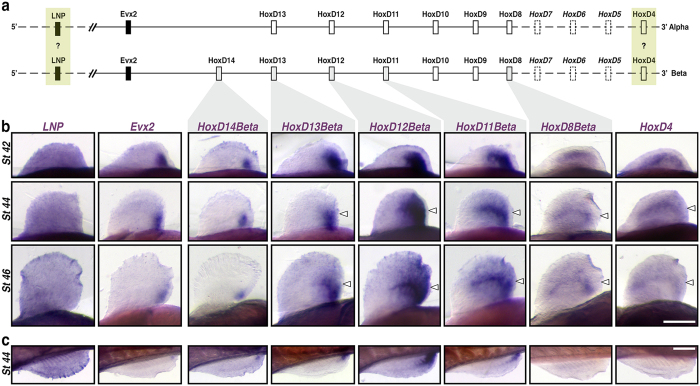
Figure 2. Expression of HoxD cluster genes in paddlefish paired fins.
(a) Schematic representation of Alpha and Beta HoxD clusters based on ref. 13. Gene key: Open boxes – Hox genes; closed boxes – non-Hox genes; solid lines – genes characterized and attributable to either Alpha and Beta clusters based on published BAC clones; yellow boxes – genes cloned but not attributable to a specific cluster; Dashed boxes – uncharacterized genes. (b) Pectoral fin whole-mount in situ hybridizations for LNP, Evx2, HoxD14Beta, HoxD13Beta, HoxD12Beta, HoxD11Beta, HoxD8Beta, and HoxD4 from stages 42 (early fin bud), 44 (onset of endoskeletal radial differentiation), and 46 (differentiated fin – onset of feeding larva)49. Pectoral fins in ventral view, anterior to the left, distal is up; Genes are shown in columns, and developmental stages in rows. Open arrowheads denote the position of distal radial formation along the A-P axis, where HoxD expression persists22 following outgrowth of the fin-fold. (c) Pelvic fin whole-mount in situ hybridizations comparable to (b) for stage 44. Pelvic fins in medial view, anterior to the left, distal is down. Scale bars = 200 nm.

