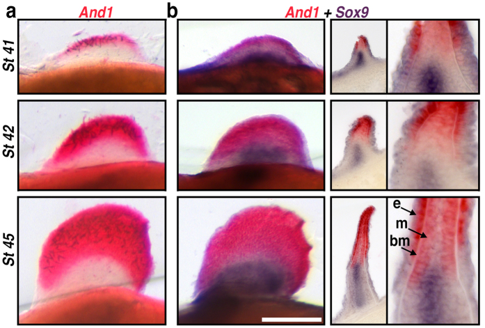
Figure 3. Actinodin1 expression in the fin-fold compartment of paddlefish.
(a,b) Pectoral fin in situ hybridizations in whole-mount, representative cross sections, and magnifications. Stages are shown in rows (a) Actinodin1 (And1), an early molecular marker for cells contributing to the fin-fold. And1 transcripts (red) appear in the presumptive distal fin and fin-fold of early fin buds (stage 41) and persist as the fin-fold elongates (stages 42 and 45). (b) Double in situs for the pre-chondrogenic marker Sox9 (purple) and And1 (red) reveal that endochondral and dermal compartments remain separate throughout fin development. Cross sections (and magnification) reveal And1 transcripts in both the fin-fold mesenchyme (m) and the ectoderm (e) adjacent to the basement membrane (bm). The slight proximo-distal overlap between And1 and Sox9 expression presages the arrangement of the dermal fin skeleton later in development. Scale bars = 200 nm.

