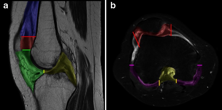
Fig. 1.
The locations at which enhancing synovial thickening on MRI of the knee is scored according to the Juvenile Arthritis MRI Scoring system (JAMRIS) [5]. On a sagittal T1-weighted sequence (a) and an axial fat-saturated T1-weighted sequence (b) of the knee the six locations are exemplified with different colours: blue suprapatellar, red patellofemoral, green infrapatellar, yellow cruciate ligaments, purple medial and lateral posterior condyle

