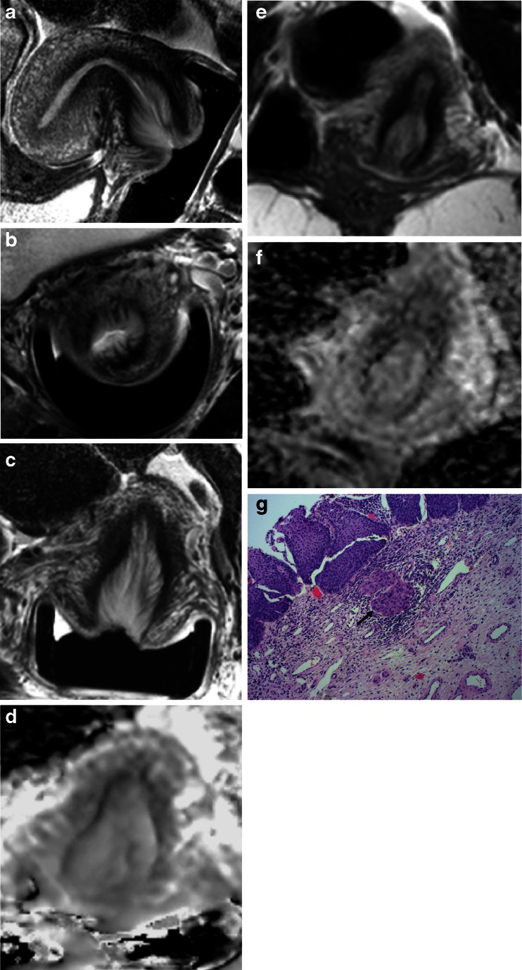
Fig. 4.
False negative on endovaginal T2-W with negative diffusion-weighted imaging (DWI) in a 27-year-old female: Sagittal (a), axial (b), coronal (c) T2-W (FSE TR 2750 ms, TE 80 ms) and ADC map (EPI, TR 6500 ms, TE 54 ms, b = 0, 100, 300, 500, 800 mm2/s, d) with corresponding external array coil T2-W coronal (FSE TR 2100 ms, TE 90 ms, e) and ADC map (EPI TR 3230 ms, TE 53 ms, b = 0, 100, 300, 500, 800 mm2/s, f). There was no evidence of tumour on MRI with either technique, but a 6-mm microinvasive tumour (Stage Ia) was noted on histopathology (g, arrow)

