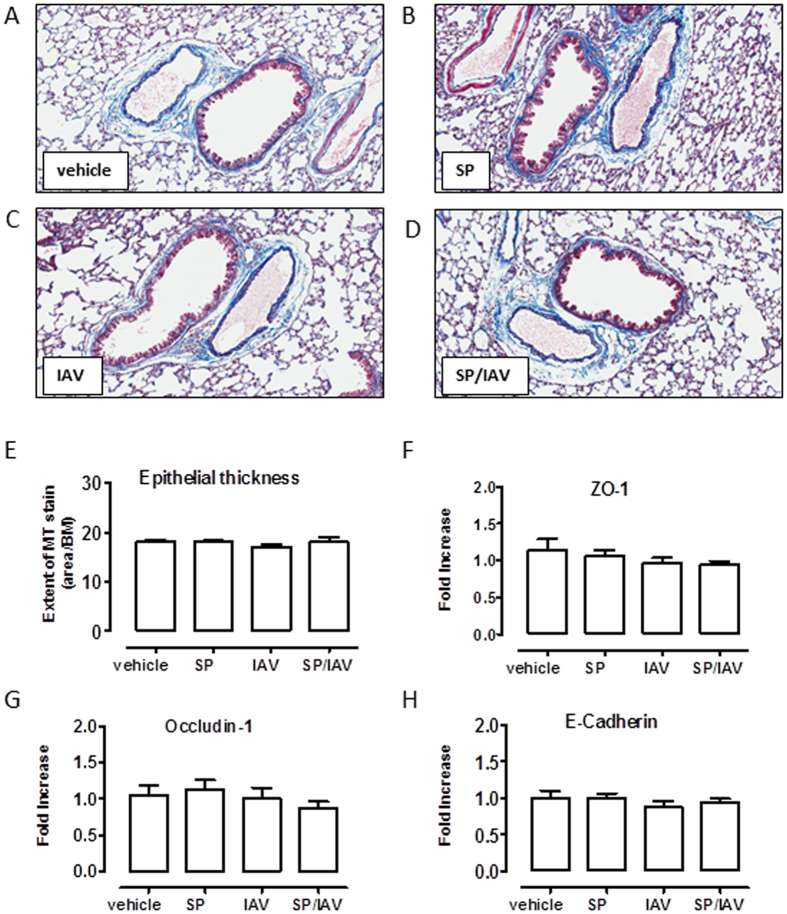
Figure 8. Neonatal co-infection is not associated with airway epithelial disruption.
Representative histological images (A–D) of Massons Trichrome (MT) staining of lung tissue from mice exposed to infection in early life. Quantification of epithelial thickness (E) is expressed as area of MT stain (purple)/basement membrane length (n = 5−11). Taqman PCR analysis of epithelial integrity markers (F) ZO-1, (G) occludin-1 and (H) E-Cadherin are expressed as fold-increase compared to vehicle-treated control and normalised to GAPDH housekeeping gene (n = 6).

