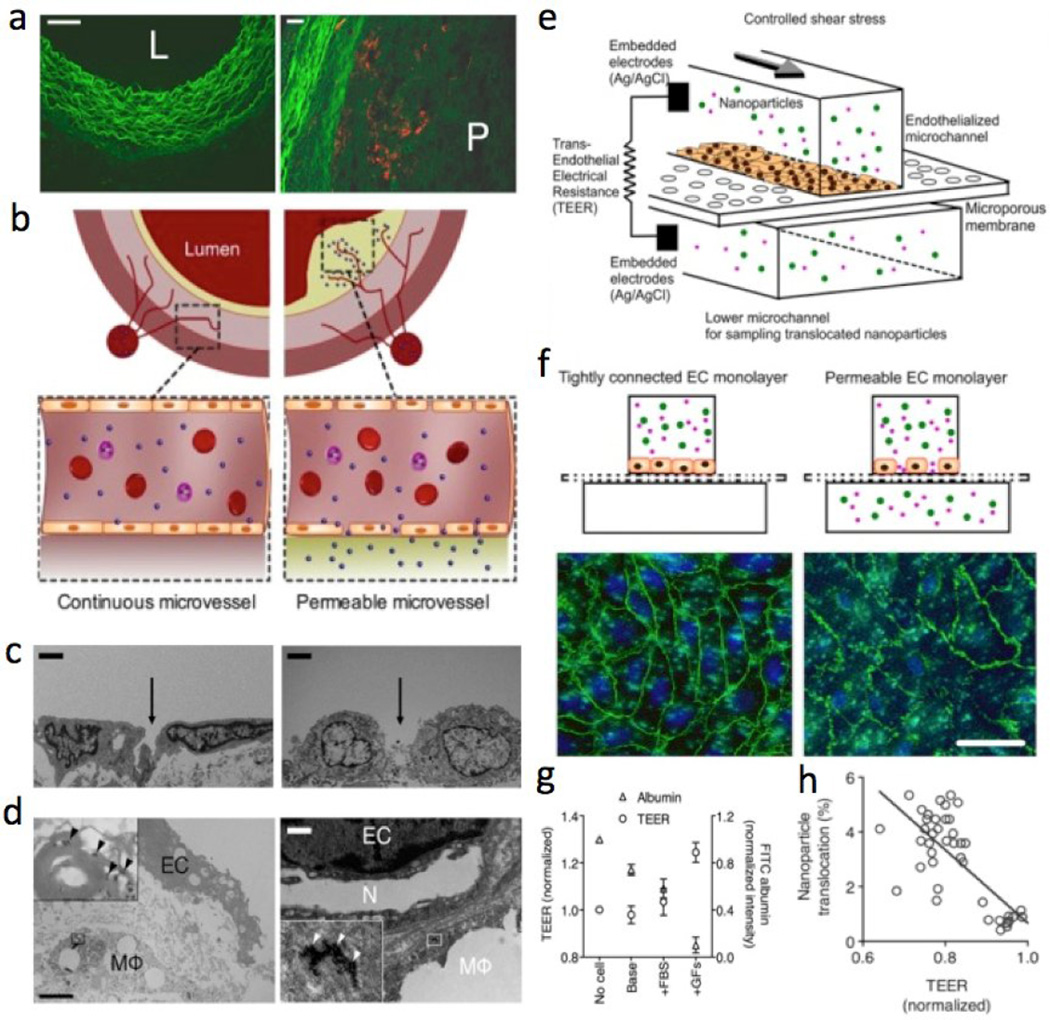
Figure 5. Probing nanoparticle translocation across the permeable endothelium using an in vitro microfluidic model with in vivo validation.
(a) Fluorescence microscopy images of typical cross-sections of the healthy wall (L, lumen) and the atherosclerotic vessel with plaque (P). (b) Cross-section schematics of continuous normal and permeable capillaries that penetrate into the atherosclerotic plaque from the vasa vasorum. (c) TEM of the endothelial lining of a normal/healthy vessel wall (left) compared to a permeable endothelial layer with a large gap (right). (Scale bar, 2µm). (d) High resolution TEM showing ECs with a gap between them and a macrophage (MΦ) behind it. At a higher magnification, multiple individual nanoparticles (arrowheads) can be found. (Scale bar, 2µm.) The neovessel (N) within the plaque is bordered with a lipid-loaded MΦ, which itself has taken up nanoparticles as well. (Scale bar, 1µm.) (e) Diagram of an endothelialized microfluidic device enabling TEER measurement across the EC layer. (f) Permeable EC layer with disrupted adherens junctions between ECs, as evidenced by patchy expression of VE-cadherin (green) in the image on the right compared to the left. Nuclei (DAPI). (Scale bar, 20µm). (g) ECs in different culture media show different permeability (shown by TEER and FITC-albumin translocation). (h) Nanoparticle translocation and TEER from these experiments are inversely correlated (r2 = 0.54, P < 0.0001). Reprinted with permission from PNAS [209].

