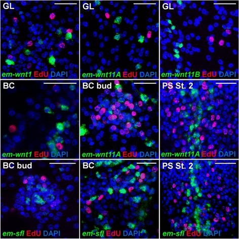
Fig. 7.

Lack of EdU incorporation in cells expressing Wnt and SFRP genes in E. multilocularis metacestodes. Green: Whole-mount in situ hybridization (WMISH); red: EdU detection; blue: nuclear DAPI staining. EdU labeling was done by culturing metacestodes in vitro for 5 hours with 50 μM EdU. Notice the lack of co-localization between WMISH and EdU for each gene at each stage. GL: Germinal layer; BC bud: Early brood capsule bud (shown from above for all genes except for em-wnt11A and em-wnt1, for which it is shown from a side); PS st 2: Second stage of protoscolex development following the system of Leducq and Gabrion [25]. Bars: 20 μm
