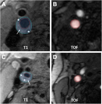
Fig. 5.

Asymptomatic (a, b) and symptomatic (c, d) internal carotid arteries (ICA) with AHA-LT V (white arrow = lipid-rich NC, white star = and small calcifications). Modified remodeling index (mRI) was calculated by dividing the outer vessel area in the T1-image (area within the blue circle, A:67.2 mm2; C:46.7 mm2) with the lumen-area in the TOF-angiography 40 mm distal to bifurcation in a normal configurated vessel segment (area within the red circle, B:24.4 mm2,D:25.7 mm2). Relatively high mRI (2.77) in asymptomatic (a, b) and relatively low mRI (1.81) in symptomatic artery
