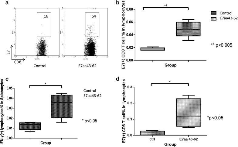
Fig. 2.

Characterization of systemic E7-specific CD8+ T cells and local E7-specific activated CD8+ T cells in tumor bearing mice. 3 × 10⁴ TC-1-Luc cells were submucosally injected into the right buccal area of C57BL/6 mice (five per group). Three days after tumor injection, mice were vaccinated intratumorally with or without 50 μg of synthetic HPV-16 E7aa 43-62 peptide for four times in a 4-day intervals. 21 days after tumor injection, peripheral blood was collected and the spleen and tumor were harvested. Cells obtained from the peripheral blood and tumor were stained with PE-conjugated HPV16 H-2D-RAHYNIVTF tetramer and APC-conjugated CD8 monoclonal antibody followed by flow cytometry analysis. Spleenocytes were stimulated with HPV16 E7aa49-57 peptide in the presence of GolgiPlug and IFN-γ-secreting CD8+ T cells were detected by intracellular cytokine staining followed by a flow cytometry analysis. a Representative flow cytometry images showing the amount of E7-specific CD8+ T cells per 1 × 10⁵ lymphocytes in the peripheral blood of various groups. b Bar graph depicting the amount of E7-specific CD8+ T cells per 1 × 10⁵ lymphocytes in the peripheral blood of various groups (mean ± SD). c Bar graph depicting the amount of IFN-γ positive lymphocytes per 1 × 10⁵ splenocytes (mean ± SD). d Bar graph depicting the percentage of E7-specific CD8+ T cells in all lymphocytes in the buccal tumor (mean ± SD)
