
Figure 2. Reduced activity of GABAA receptors is involve in soluble Aβ-mediated impairment of LTP in the CA1 region of hippocampus.
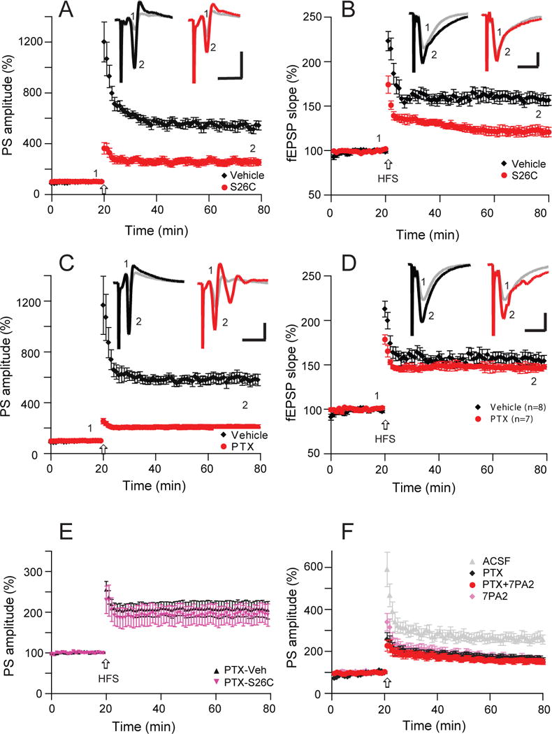
(A) Synthetic Aβ40S26C dimer significantly reduces population spike LTP (red, n=6) induced by high-frequency stimulation (HFS, arrow) in comparison with the vehicle effect on LTP (black, n=7). (B) Synthetic Aβ40S26C dimer also inhibited simultaneously recorded fEPSP-LTP (red), while the vehicle had no effect on this regular LTP (black). (C) A GABAA antagonist (picrotoxin, 50 μM) significantly reduce population spike LTP (red, n=8). (D) The same dose of GABAA antagonist (picrotoxin, 50 μM) has no effect on the simultaneously recorded usual field EPSP (blue, n=8). (E) Hippocampal population spike-LTP was also reduced by the GABAA receptor antagonist, picrotoxin (50 μM) (black, n=7), while pretreatment with picrotoxin prevented the S26C-dimer induced further decrease of PS-LTP (red, n=6). (F) A low dose of a GABAA antagonist (picrotoxin, 10 μM) occluded 7PA2 CM-mediated impairment of population spike LTP (red, n=8). Inset traces are typical population spikes (A, C) and field excitatory postsynaptic potentials (fEPSPs) (B, D) recorded before (gray) and after (black or red) HFS for each condition. Horizontal calibration bars: 10 ms; vertical bars: 2 mV (A, C) or 1 mV (B, D).