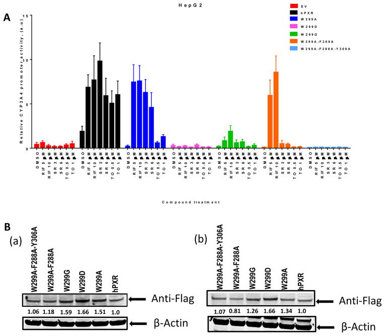
Figure 2. The activity and expression of WT hPXR and hPXR mutants in HepG2 cells.
(A) HepG2 cells transiently transfected with empty vector (EV), WT hPXR (hPXR) or hPXR mutant as indicated, CYP3A4-luc, and CMV-Renilla were treated with DMSO or different concentrations of agonists as indicated for 24 h prior to luciferase assay. (B) Western blot showing Flag-PXR (WT or mutants) protein levels in HepG2 cells upon (a) RIF [5 μM] and (b) DMSO treatment. The numbers below the protein bands indicate the relative intensity of the protein bands, with the wild-type Flag-PXR sample set as “1”. Anti-Flag antibody was used to detect Flag-PXR. Anti-β-actin was used to detect β-actin (as loading control).

