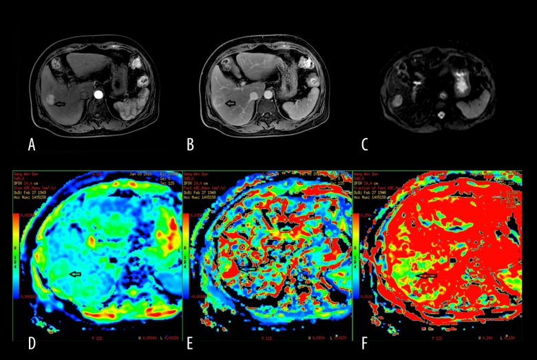
Figure 3.
A 63-year-old male man with primary hepatocellular carcinoma. (A) Contrast-enhanced DWI during the arterial phase showing lesion enhancement in segment IV of the liver. (B) The lesion enhancement degree in the delayed phase decreases obviously compared to that of the arterial phase. (C) The signal of the lesion is slightly higher than hepatic parenchyma when b=0 s/mm2. (D) Mapping of the estimated value of the D parameter. The average value in the lesion ROI was D=1.19×10⁻³ mm2/s. The signal of the lesion is slightly lower than hepatic parenchyma. (E) Mapping of the estimated value of the D* parameter. The average value in the lesion ROI was D*=22.4×10⁻³ mm2/s. The signal of the lesion is slightly lower than hepatic parenchyma. (F) Mapping of the perfusion-related diffusion fraction (f) with a value of 12.2%.

