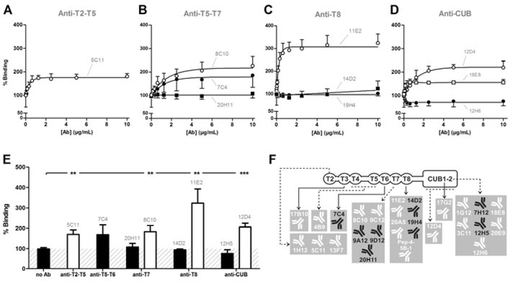
Fig. 5. Anti-T2-CUB2 mAbs stimulate the binding of ADAMTS13 to folded VWF.
Folded pVWF was captured on the anti-VWF A1 domain mAb 6D1. Recombinant ADAMTS13, pre-incubated with the respective mAb (½ dilution series), was added to captured pVWF and detected with biotinylated anti-MDTCS mAbs and HRP-labeled streptavidin. The absence of mAb (‘no Ab’) was set as 100% binding. Error bars represent the SD of at least three independent experiments. Non-functional mAbs 7C4 and 20H11 (B), 14D2 and 19H4 (C) and 12H5 (D) are representative for all non-functional anti-T5-T6, anti-T7, anti-T8 and anti-CUB mAbs, respectively. Likewise, activating mAbs 5C11 (A), 8C10 (B), 11E2 (C), 12D4 and 18E8 (D) are representative for all activating anti-T2-T5, anti-T7, anti-T8 and anti-CUB mAbs, respectively. (E) The percentage binding induced by each anti-T2-CUB2 mAb at 10 µg/mL was calculated and compared with the percentage binding in the absence of the mAb, using the Student t-test. In line with A–D, mAbs 7C4, 20H11, 14D2 and 12H5 represent respectively the non-functional anti-T5-T6, anti-T7, anti-T8 and anti-CUB mAbs. In addition, all functional anti-T2-T5, anti-T7, anti-T8 and anti-CUB mAbs are represented by mAbs 5C11, 8C10, 11E2 and 12D4 respectively. Error bars represent the SD of three independently performed experiments. (F) Overview of the functional effect of all anti-T2-CUB2 mAbs (for details, see Table S2). Non-functional and activating mAbs are represented in black and white, respectively.

