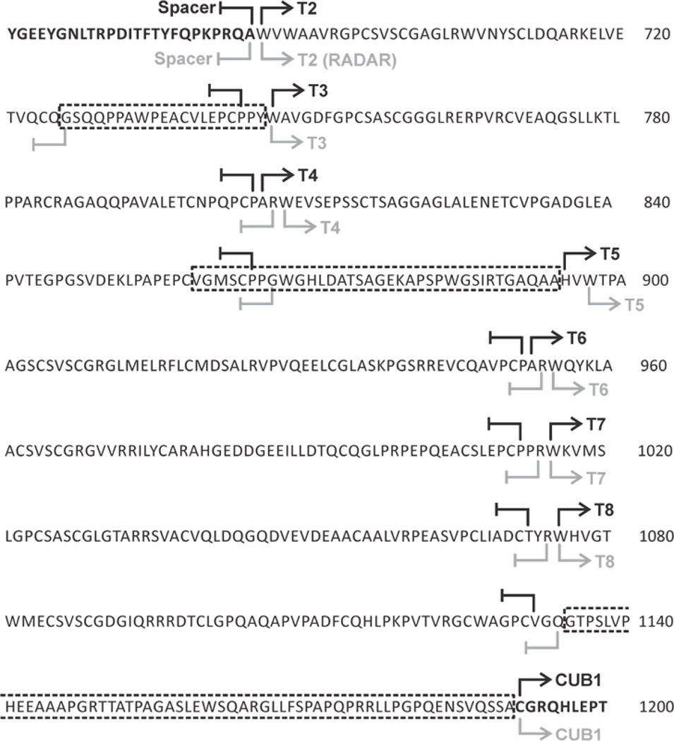
Fig. 6. The distal part of ADAMTS13 contains three linker regions.
The amino acid sequence of the distal domains is represented. Linker regions (framed with dotted lines) are located between the T2 and T3 domains (L1), the T4 and T5 domains (L2), and the T8 and CUB1 domains (L3). The current boundaries, defined by Zheng et al. [38], are represented in black and the boundaries defined by RADAR analysis are represented in gray.

