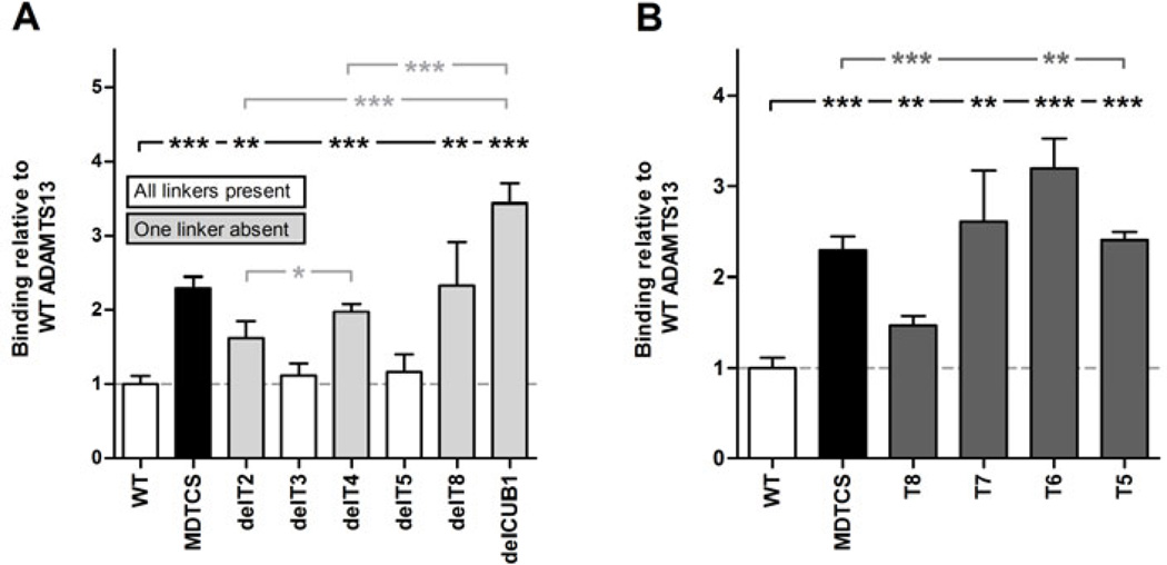
Fig. 7. Exposure of the 6A6 cryptic epitope by removal of linker regions between the distal domains or deletion of T8-CUB2 domains.
Binding of rADAMTS13 and its variants to anti-metalloprotease mAb 6A6 was tested in ELISA. Binding was detected with the anti-V5-HRP mAb and calculated relative to the binding of rADAMTS13. (A) Binding of rADAMTS13, MDTCS and the T2-CUB2 deletion variants to 6A6. White bars represent rADAMTS13 and its variants which contain all three linker regions (L1, L2 and L3), while gray bars represent rADAMTS13 variants devoid of one of the three linker regions (see Fig. 1). (B) Binding of rADAMTS13, MDTCS and the T2-CUB2 truncation variants to 6A6. Differences were statistically analysed using the Student t-test. Error bars represent the SD of at least three independent experiments.

