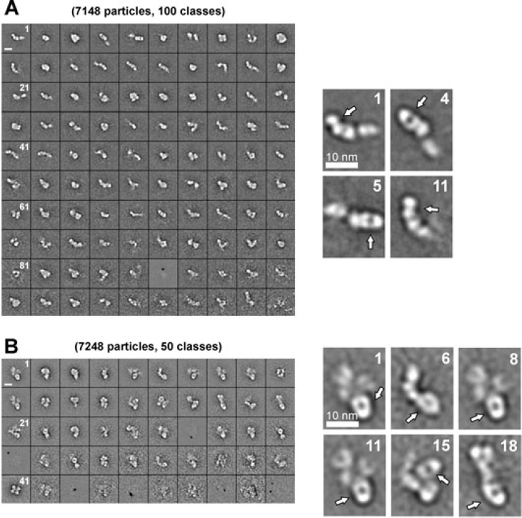
Fig. 8. EM analysis reveals flexibility around the T2 and metalloprotease domain.
EM images of purified rADAMTS13, pre-incubated with anti-metalloprotease domain 3H9 Fab (A) or anti-T2 5C11 Fab (B), were made and respectively 100 and 50 class averages of the different structures of ADAMTS13 were selected. For both conditions, magnified class averages are shown. The respective Fab fragment was easily identified due to its characteristic round shape (indicated with the white arrow). (A) Domains C-terminal from the metalloprotease domain were difficult to identify, icating flexibility around this domain. (B) Domains N- and C-terminal from T2 are visible but could not be identified, also indicating flexibility around the T2 domain. Scale bars, 10 nm.

