Figure 3.
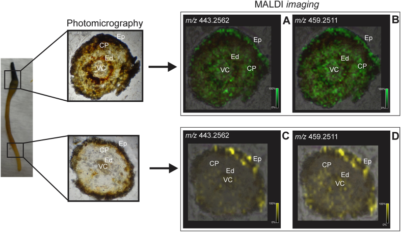
Transverse sections of root tissues from Peritassa laevigata obtained from different parts (root cap, differentiating region, and primary structure) and their MALDI-MS images reconstructed with ions m/z 443.2562 [M + Na]+ (A,C) and 459.2511 [M + Na]+ (B,D) corresponding to maytenin (1) and 22β-hydroxy-maytenin (2), respectively. CP: cortical parenchyma; Ed: endoderm; Ep: epiderm; VC: vascular cylinder.

