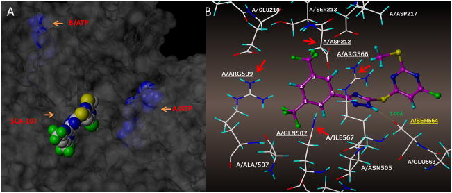
Figure 5. Molecular modeling of 7b (SCA-107) binding site in E. coli SecA. 6A.
Docking was done in SYBYL 2.0 using PDB (2FSG) with SecA dimers A and B. 7b binds to the interface of the A and B monomers, closer to A (6A), partially blocking the entrance to the ATP site. 6B: SCA-107 surrounded by amino acid residues.

