
Figure 5. c-Abl Inhibition Hypersensitizes Prostate Cancer and RCC Cell Lines to Hsp90 Inhibitors.
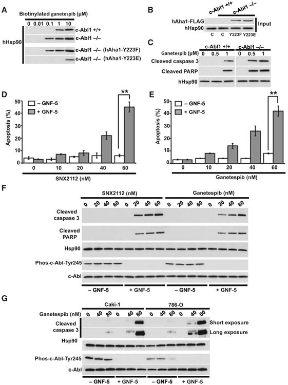
(A) Lysates from WT (c-Abl+/+) and c-Abl deficient (c-Abl−/−) MEF cell lines also carrying indicated hAha1 phospho mutant plasmids were incubated with indicated amounts of biotinylated ganetespib, followed by streptavidin agarose beads. The hHsp90α/β was detected by immunoblotting with anti-hHsp90 monoclonal antibody (16F1).
(B) Total lysate from samples in (A) were tested for hHsp90 by immunoblotting with anti-hHsp90 (16F1) and anti-FLAG antibodies.
(C) WT (c-Abl+/+) and c-Abl deficient (c-Abl−/−) MEF cell lines were treated with indicated amounts of ganetespib for 6 hr. The cleaved PARP and cleaved caspase-3 were detected by immunoblotting. The hHsp90 was used as a loading control.
(D and E) Prostate cancer PC3 cells were treated with 5 μM c-Abl inhibitor GNF-5 for 24 hr. The cells were then treated with indicated concentrations of Hsp90 inhibitors SNX2112 and (E) ganetespib for an additional 24 hr. Apoptosis was detected by FACS analysis. The errors bars in (D) and (E) represent the SD of three independent experiments (**p < 0.005).
(F) PC3 cells were treated with 5 μM c-Abl inhibitor GNF-5 for 24 hr and then with indicated concentrations of SNX2112 and ganetespib for an additional 24 hr. Hsp90, c-Abl, active c-Abl (phospho-Y245), and apoptosis indicator cleaved caspase-3 and cleaved PARP were detected by immunoblotting.
(G) RCC Caki-1 and 786-O cell lines were treated with 5 μM c-Abl inhibitor GNF-5 for 24 hr and then with indicated concentrations of SNX2112 and ganetespib for an additional 24 hr. Hsp90, c-Abl, active c-Abl (phospho-Y245), and apoptosis indicators cleaved caspase-3 and cleaved PARP were detected by immunoblotting.