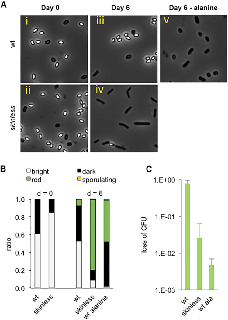
Figure 4. Consequences of Spontaneous Germination in Favorable and Unfavorable Environments.
(A) Spontaneous germination of wild-type and skinless spores in liquid mISP4. Spores were generated by exhaustion, re-suspended in mISP4 with xylose, and incubated at room temperature. Samples taken at day 0 and day 6 following initiation of incubation were analyzed by light microscopy.
(B) Quantification of (A) with fraction of phase-bright spores (white), phase-dark (black, considered to be less dormant than phase-bright spores) cells, vegetative cells, rods (green), and sporulating cells (yellow), as determined by observation using phase-contrast microscopy.
(C) Killing of skinless spores. Following incubation in mISP4 + xylose for 6 days, wild-type and skinless spores were subjected simultaneously to chloramphenicol (10 µg/ml), ampicillin (100 µg/ml), and kanamycin (50 µg/ml) for 120 min and plated on mISP4 to determine CFUs. As a control, wild-type spores were incubated with 10 mM alanine for 60 min before exposure to antibiotics. The ratio of CFUs before and after treatment with antibiotics “loss of CFU” is reported. For wild-type versus skinless, p = 0.03; for wild-type versus wild-type + alanine, p = 0.03 (unpaired t test, two tailed). Shown is the mean, and error bars represent the SD.

