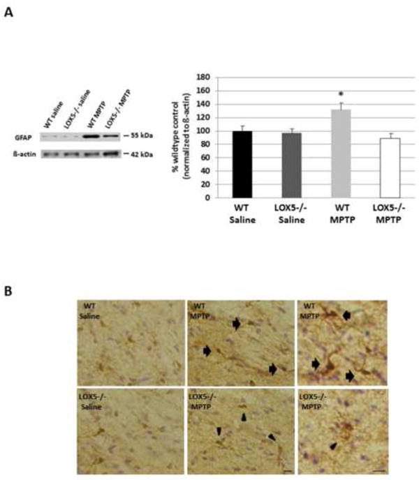
Fig. 5. 5-LOX isozyme effects on inflammatory markers following toxic insult.

(A) The astrocytic protein GFAP was semi-quantitatively assessed using Western blot analyses of striatal homogenate from WT and null 5-LOX littermates treated with saline or MPTP (n=6-8/group). Immunoreactivity for GFAP was measured by optical density and normalized to the housekeeper β-actin. Data are shown as means±SEM. * P<0.05. (B) Immunohistochemical staining for the microglial protein Iba-1 was visualized using the brown chromogen DAB with counterstaining by cresyl violet. Coronal brain sections containing substantia nigra from WT and null 5-LOX littermates treated with saline or MPTP were utilized. Panel on far right shows microglial morphology: arrows indicate rounded cell bodies with short, thick processes; arrowheads denote ramified cell bodies with long, branching processes. Bar = 25 μm.
