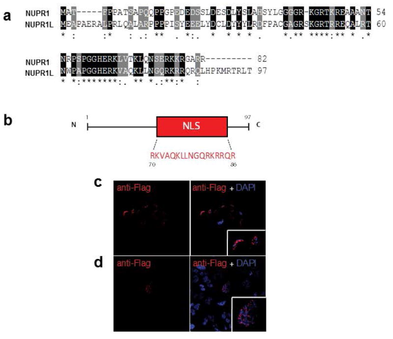
Figure 2. Primary structure analysis and nuclear localization of Nup1L protein.

(a) Pairwise alignment of the Nupr1 and Nupr1L proteins displaying a 60 % of homology. Residues labeled with a (*) indicate 100% identity, residues labeled with (:) indicate positions with conservative substitutions, residues labeled with (.) indicate substitutions less conservative. (b) Schematic representation of Nupr1L protein, which contains a Nuclear Localization Signal (NLS). The Flag-Nupr1L immunofluorescence in MiaPaca-2 (c) and Hela cells (d) reveals its nuclear localization. The cells were transduced with a lentiviral vector (6His-Flag-Nupr1L) and were plated on coverslips. The mouse Flag was revelated using Alexa fluorantimouse IgG488. Nuclei are stained by blue fluorescent DAPI.
