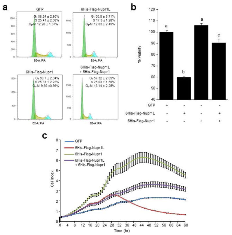
Figure 8. Nupr1L decreases the pancreatic cancer cells viability.

MiaPaca-2 cells were infected by 6His-Flag-Nupr1L and 6His-Flag-Nupr1 lentiviral vector, individually and together. GFP overexpressing cells were used like control (e) Cytometric cell cycle analysis showing a G1 cell cycle arrest with a significant decrease of S phase induced by Nupr1L overexpression and abolished by the concomitant overexpression of Nupr1. (f) Cell viability measured by Cell Titer-Blue assay. Decrease of viability is showed in cells infected by 6His-Flag-Nupr1L vector but not in infected cells byboth lentiviral vectors, 6His-Flag-Nupr1 and 6His-Flag-Nupr1L. Cell viability in GFP infected cells was normalized to 100%. Values are expressed as mean ± S.D. Means sharing the same superscript letter are not significantly different from one another (P< 0.05). (c) Real-time cell proliferation assay using electric impedance as a measure of cell proliferation. Electrical impedance was normalized according to the background measurement at time point 0. Impedance measurements were carried out for 72 h. Data points represent mean values ± S.D. (n = 3). Results show lower rate of proliferation in NUPR1L-overexpressing cells than GFP-transduced cells. The concomitant overexpression of Nupr1 abolished the effect induced by Nupr1L on the proliferation cell, reaching intermediate values to both infected cells (6His-Flag-Nupr1L and 6His-Flag-Nupr1 cells).
