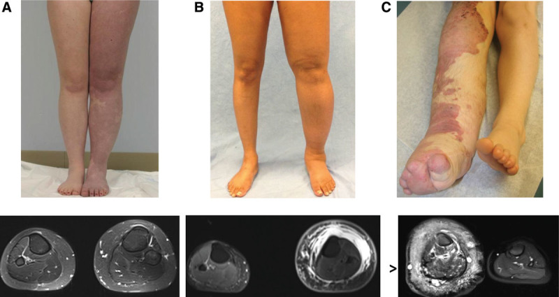
Fig. 1.

Vascular malformations with phenotypical similarities to lymphedema–capillary malformation. A, Adult female with diffuse capillary malformation with overgrowth (DCMO) who had an epiphysiodesis of the left lower extremity. MRI illustrates increased subcutaneous adipose deposition with venous phlebectasia. B, Adult female with primary lymphedema of the left leg. MRI shows subcutaneous adipose hypertrophy and edema. C, Patient with Klippel–Trenaunay syndrome of the lower extremity. MRI indicates vascular malformations affecting all structures of the limb. The subcutaneous tissue contains microcystic lymphatic malformation, a persistent embryonal vein laterally (marginal vein of Servelle), and enlarged saphenous veins medially. The subfascial compartment contains venous malformations of the muscles and bone. Arrowhead identifies the marginal vein of Servelle.
