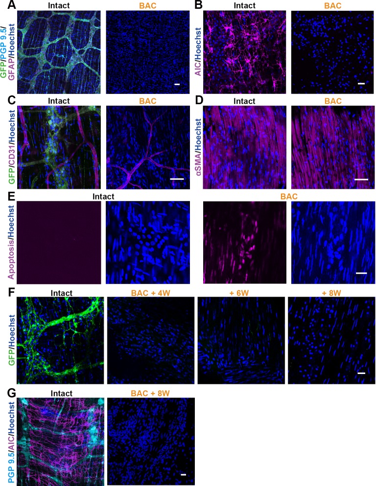
Fig 3. The Effects of BAC Treatment Confirmed by Immunostaining.
Two weeks after BAC treatment, immunohistochemically evaluated the GFP-positive NCCs and various markers in the lesion sites. (A-D) NCCs stained with GFP antibody was entirely deleted from the BAC treated colon (A). Not only PGP 9.5-positive enteric ganglion cells, but also GFAP-positive glial cells were disappeared (A). Additionally, pacemaker cells, known as interstitial cells of Cajal (B), were also absent from the lesion sites. (C, D) No obvious defects were observed with the CD31-positive endothelial cells (C) and the αSMA-positive smooth muscle cells (D) in the ablated area. (E) Not in the intact colon, but in the BAC treated colon, the apoptosis marker was frequently detected by the ApoTag kit one day after BAC treatment. (F) Time course dependent change of the ablated segment was demonstrated that the enteric neural networks were irreversibly absent for 8 weeks after the BAC application. (G) At this time point (8 weeks), ICC was also irreversibly absent, similar to the case in the ENS networks. Scale: (A-D, F) 50 μm, (E, G) 20 μm

