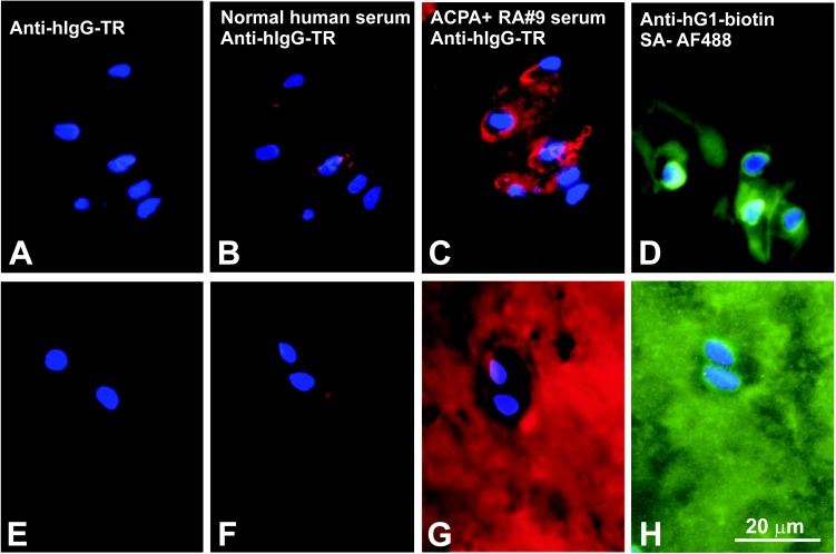
Fig 4. Immunohistochemical localization of ACPA-reactive (citrullinated) epitopes in OA and RA cartilage sections.
(A-D) Frozen sections of OA knee (tibial plateau) cartilage were immunostained with (A) Texas red-labeled anti-human IgG (anti-hIgG-TR), (B) normal (ACPA-negative) human serum followed by anti-hIgG-TR, (C) ACPA+ serum (RA#9) followed by anti-hIgG-TR, or (D) a biotinylated anti-human G1 antibody followed by Alexa Fluor 488-labeled streptavidin (SA-AF488). (E-H) Frozen sections prepared from the RA tibial plateau cartilage were immunostained with the same sera and antibodies as listed for A-D. Cell nuclei in all sections were visualized by DAPI staining. (A and E) Anti-hIgG-TR alone did not stain the sections, and (B and F) negligible reaction (red fluorescence) was observed when the tissues were first stained with ACPA- serum. (C) The ACPA+ serum primarily stained the chondrocyte pericellular matrix in the OA cartilage, but (G) it diffusely stained the entire matrix of the RA cartilage. Similar staining patters to those with ACPA+ serum were observed when (D) the OA and (H) RA cartilage sections were incubated with biotinylated anti-hG1 mAb (green fluorescence), suggesting at least partial co-localization of PG G1 and citrullinated epitopes in both OA and RA cartilage.

