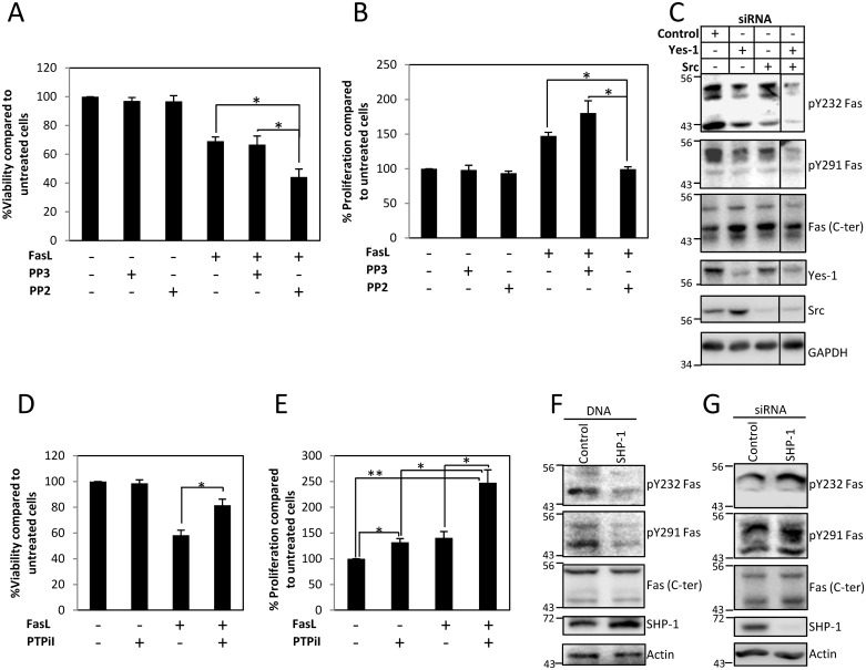
Fig 6. Fas death domain tyrosine phosphorylation is regulated by Src family kinases (SFKs), Src and Yes-1, and protein tyrosine phosphatase SHP-1.
(A) SW480 cells expressing V5-tagged Fas protein were pretreated with vehicle (DMSO), 10 μM PP3 (negative control for PP2), or 10 μM PP2 for 30 min and then incubated with 10 ng/ml FasL crosslinked with M2 for 24 h before subjected to cell viability analysis using WST-1 assay. Percentages of viability compared to untreated control cells are shown as means ± SEM from three independent experiments (* p < 0.05, unpaired t test). (B) SW480 cells were synchronized to G1 phase by serum deprivation for 24 h and then treated with vehicle (DMSO), 10 μM PP3 (negative control for PP2), or 10 μM PP2 for 30 min, followed by 0.1 ng/ml of sFasL for 4 h before analyzing the proliferation by BrdU incorporation measurement using microplate-based method. Means ± SEM of three independent experiments are shown (* p < 0.05, unpaired t test). (C) SW480 cells were transiently transfected with control siRNA or siRNA against Src, Yes-1, or Src and Yes-1 for 72 h. Cell lysates were then collected and subjected to SDS-PAGE and immunoblotting with indicated antibodies. (D) SW480 cells expressing V5-tagged Fas protein were pretreated with vehicle (DMSO) or 50 μM PTPiI for 30 min and then incubated with 10 ng/ml FasL crosslinked with M2 for 24 h before being subjected to cell viability analysis using WST-1 assay. Percentage of viability compared to untreated control cells are shown as means ± SEM from three independent experiments (* p < 0.05, unpaired t test). (E) SW480 cells were synchronized to G1 phase by serum deprivation for 24 h and then treated with vehicle (DMSO) or 50 μM PTPiI for 30 min followed by 0.1 ng/ml of sFasL for 1 h before analyzing the proliferation by BrdU incorporation measurement using microplate-based method. Means ± SEM of three independent experiments are shown (* p < 0.05, ** p < 0.01, unpaired t test). (F) SW480 cells were transfected with control vector or SHP-1 for 24 h and (G) with control siRNA or siRNA against SHP-1 for 48 h, then synchronized in G1 phase by serum deprivation for 24 h before treatment with 0.1 ng/ml FasL for 5 min. Cell lysates were then collected and subjected to SDS-PAGE and immunoblotting with indicated antibodies. The specificity of anti-pY232 and anti-pY291 Fas antibodies is demonstrated in S11 Fig. Numerical values underlying the data summary displayed in this figure can be found in S1 Data.

