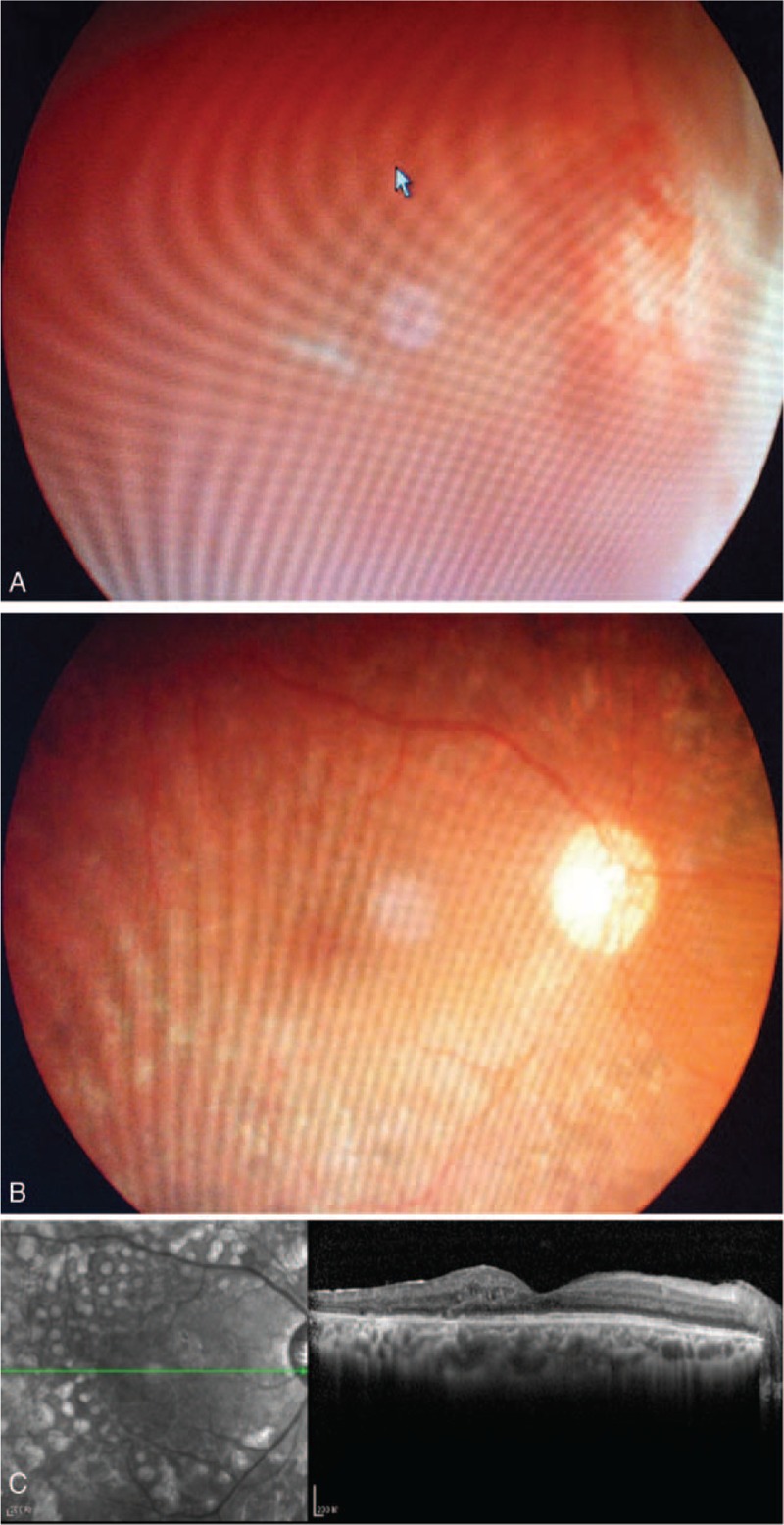
FIGURE 2.

(A) A 30-year-old man with diagnosis of type I diabetes for 1 year showing neovascularization in front of the optic disc, and ultrasonography showed retinal detachment. He received Lucentis 1 week before vitrectomy. (B) One and a half year after surgery, his retina remained attached without neovascularization, and his BCVA was 20/60. (C) The OCT image of macular area at 1.5-year follow-up.BCVA = best-corrected visual acuity, OCT = optical coherence tomography.
