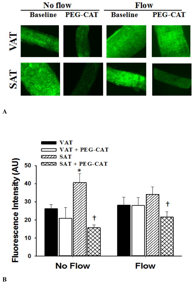
Figure 7.
Hydrogen peroxide (H2O2) production in visceral (VAT) and subcutaneous adipose tissue (SAT) arterioles. Fluorescence detection of H2O2 assessed by dichlorodehydrofluorescein diacetate (DCF-DA; 2 μM) was determined in the presence and absence of PEG-CAT under no flow and after 30 minutes of intraluminal flow (pressure gradient of 60 cmH2O). Panel A shows representative images of SAT and VAT vessels under no flow and flow conditions and in the presence and absence of (PEG-CAT, 500 U/ml). During flow, the presence of PEG-CAT significantly reduced DCF-DA fluorescence in SAT arterioles, but had no effect on DCF-DA fluorescence in VAT arterioles. There was no effect of flow on H2O2 production compared to baseline. Panel B shows the summarized data describing the fluorescence intensity in vessels with and without flow. Data are presented as mean ± SEM. *P<0.05 vs. visceral fat without flow. †P<0.01 vs. baseline without PEG-CAT.

