Fig. 4.
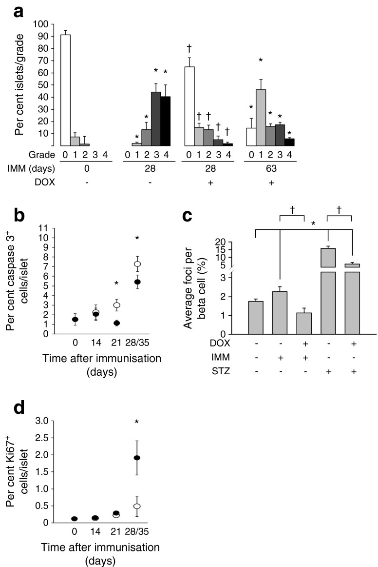
BPTL mice overexpressing PAX4 display reduced insulitis, correlating with decreased islet apoptosis and DNA damage and increased proliferation. (a) Insulitis was scored as grade 0–4 according to the percentage of infiltrated islet area (0: 0%; 1: <10%; 2: >10% and <55%; 3: >55% and <75%; 4: >75%). n = 5–6, *p < 0.05 vs immunised day 0 and (−) DOX group; † p < 0.05 vs immunised day 28 and (−) DOX group. (b) Apoptosis was assessed by immunohistochemical analysis of cleaved caspase-3 in pancreatic islets from BPTL mice treated (black ovals) or not (white ovals) with DOX and killed at the time points. As similar results were obtained for days 28 and 35, the data were combined as a single time point. n = 5–6, *p < 0.05, (+) vs (−) DOX groups within each time point. (c) The average number of 53BP1 foci per beta cell was assessed from pancreatic islets of either 14-day-old immunised BPTL mice or 2-day-old STZ-treated PAX4 mice exposed (+) or not (−) to DOX. n = 5–6, *p < 0.05 vs untreated mice; † p < 0.05 vs DOX-untreated immunised or STZ-treated mice. (d) Cell proliferation was assessed by the combined immunohistochemical analysis of Ki67 in pancreatic islets from BPTL mice treated (black ovals) or not (white ovals) with DOX and killed at the time points. n = 5–6, *p < 0.05, (+) vs (−) DOX groups within the time point. IMM, immunised

