
Figure 6. Examples of optical chemical probes being used in animal models for noninvasive cancer diagnostics, in vivo, and ex vivo surgical guidance.
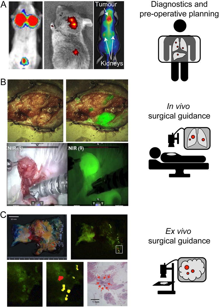
A. LEFT: 6QC (20 nmol IV), imaged after 4 hr in 4T1 Breast cancer mouse model, MIDDLE: Folate-Dylight680 (10 nmol IV), imaged after 4 hr in FR-expressing L1210A tumor mouse model, RIGHT: C-SNAF (5 nmol IV), imaged after 1 hr in three times DOX treated tumor-bearing mice. B. TOP: CTX:Cy5.5. Image shows white light and white light with NIR overlay on a canine tumor tissue sample. BOTTOM: 6QCNIR injected 6h prior to surgery. Image shows white light on the left and fluorescence using the da Vinci® surgical system on right. C. gGlu-HMRG (3 mL of 5 μM in 0.5% v/v DMSO in RPMI1640) topically applied to patient specimen diagnosed with invasive ductal carcinoma (papillotubular) and imaged after 5 min.