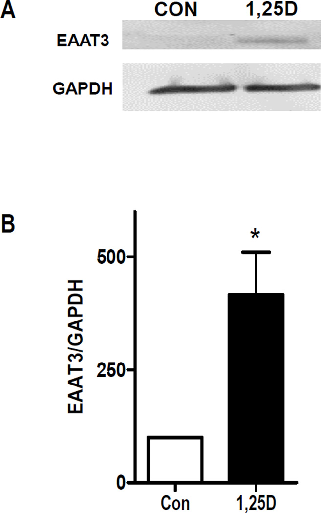
Figure 2. Expression of EAAT3 in 1,25D-treated hTERT-HME1 cells.
A, hTERT-HME1 cells were treated with 100 nM 1,25D or ethanol vehicle (Con) for 48 h. Whole cell lysates were analyzed by Western blotting with antibodies directed against EAAT3 or GAPDH. Blot represents one of three biological replicates. B, Quantitation of western blot data. Each bar represents mean ± SD of triplicates. *p <0.05 as measured by one-tailed, unpaired student t-test.

