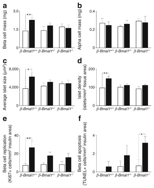
Fig. 6.
Effects of adult beta cell-specific Bmal1 deletion on beta cell mass, proliferation and apoptosis in vivo following exposure to either chow or HFD. Bar graphs depicting (a) beta cell mass, (b) alpha cell mass, (c) average islet size, (d) islet density, (e) frequency of beta cell replication and (f) apoptosis in β-Bmal1+/+, β-Bmal1+/− and β-Bmal1−/−mice exposed to 8 weeks of either chow (white bars) or HFD (black bars). (a–f) Graphs represent mean ± SEM (n=5–7 per group). Statistical significance is denoted by *p<0.05, and **p<0.01

