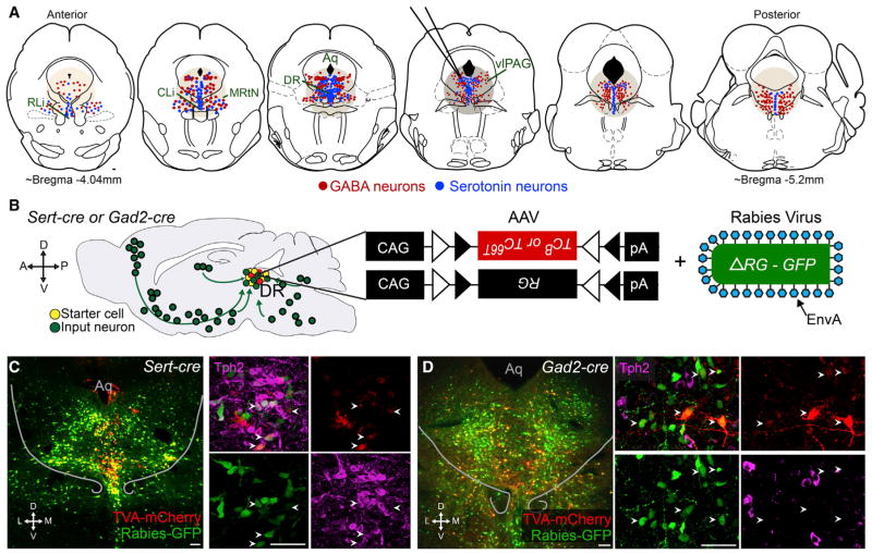
Figure 1. DR Serotonin and GABA Neurons as Starter Cells for Rabies-Based Transsynaptic Tracing.
(A) Schematic representation of serotonin (blue) and GABA (red) neurons on coronal sections through the DR and surrounding regions, including the central and rostral linear nucleus raphe (CLi and RLi, respecitively), midbrain reticular nucleus (MRtN), and ventrolateral PAG (vlPAG). The approximate location targeted for viral injections and spread of infection is indicated with tan circles. Only serotonin and GABA neurons within these regions are drawn. Aqueduct (Aq).
(B) Schematic of rabies-based transsynaptic tracing. Sert-cre or Gad2-cre mice were transduced with two AAVs in the DR followed by EnvA-pseudotyped, glycoprotein (RG)-deleted, and GFP-expressing rabies virus. Serotonin or GABA starter cells are labeled in yellow, and presynaptic partners throughout the brain are labeled in green, as shown on a schematic sagittal section of the mouse brain. TCB, wild-type TVA-mCherry fusion used in Figures 2–5; TC66T, TVA-mCherry with a point mutation (66T) in the TVA receptor used in Figure 7; CAG, a ubiquitous promoter; triangles: loxP and Lox2272 sites that cause the transgene expression to be Cre dependent (FLEx).
(C) Left, 60 μm coronal section through the DR of a Sert-cre tracing brain showing the location of starter cells (yellow). Right, z projected confocal stacks of a different Sert-cre tracing brain in approximately the same position, triple labeled in green for GFP from rabies virus, in red for mCherry from TCB, and in magenta with anti-Tph2 staining. All starter cells are Tph2 positive (arrowheads).
(D) Same as in (C), except from Gad2-cre tracing. Right panels show that none of the starter cells (arrowheads) are Tph2 positive.
Scale, 100 μm. In this and all other figures, abbreviations are as follows: A, anterior; P, posterior; D, dorsal; V, ventral; M, medial; L, lateral. Anatomical schematics and coordinates here and throughout are modified from Paxinos and Franklin (2001). Figure S1 describes further characterization of starter cell populations and the rabies tracing technique.

