
Fig. 3.
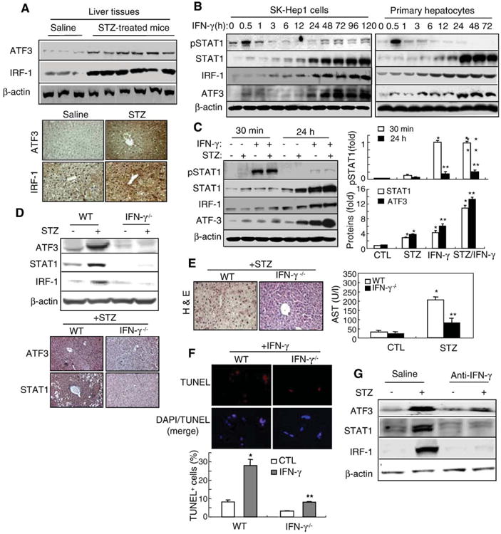
ATF3 expression induced by STZ or IFN-γ was abolished in IFN-γ−/− mice. (A) ATF3 and IRF-1 protein expression are enhanced in STZ-injected mice. Western blot analysis (upper) and immunostaining (×100) (lower). (B) SK-Hep1 and primary hepatocytes were treated with IFN-γ for the indicated time points and then subject to immunoblotting. (C) Primary hepatocytes were treated with IFN-γ and/or STZ for 30 min or 24 h and then subjected to Western blot analysis (left). Relative expression of pSTAT1, STAT1, and ATF3 was quantified (right). Values represent means±S.E.M. from 3 independent experiments. *P<0.05, **P<0.01 in comparison with corresponding control groups. (D) ATF3 and IRF-1 protein expression in STZ-injected wild-type and IFN-γ−/− mice. Western blot analysis (upper) and immunostaining (×100) (lower). (E) H&E staining (left) and AST levels (right) in STZ-treated wild-type and IFN-γ−/− mice. *P<0.01, **P<0.05 in comparison with corresponding non-treated control groups. (F) Isolated primary hepatocytes were treated with IFN-γ and then DAPI staining and TUNEL analyses were performed (upper). TUNEL+-cells were quantified (lower). Values represent means±S.E.M. from 3 independent experiments. *P<0.01, **P<0.05 in comparison with corresponding non-treated control groups. (G) Mice were injected with STZ (60 mg/kg, i.p.) and anti-IFN-γ Ab (2 μg, i.v.). Western blot analysis was performed.