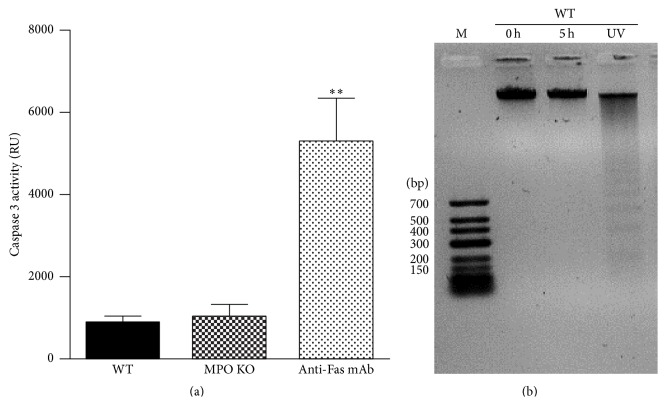
Figure 9.
Determination of caspase 3 activity and DNA fragmentation. (a) Freshly isolated nucleated cells or cells 5 h after isolation (5 × 10⁶ cells/mL) from BALF of WT and MPO KO mice treated for 36 h by intranasal application of LPS (0.3 mg/kg) were lysed and caspase 3 activity was determined by enzymatic assay. Cells treated with anti-Fas antibody were used as a positive control. Values represent mean ± SEM from 8–10 mice with significant difference between WT or MPO KO mice and positive control (∗∗ p < 0.01). (b) DNA extracted from freshly isolated cells (0 h) or cells incubated in vitro for 5 h (5 h). Cells were isolated from BALF of WT mice treated for 36 h by intranasal application of LPS (0.3 mg/kg). UV-irradiated BALF cells were used as a positive control.

