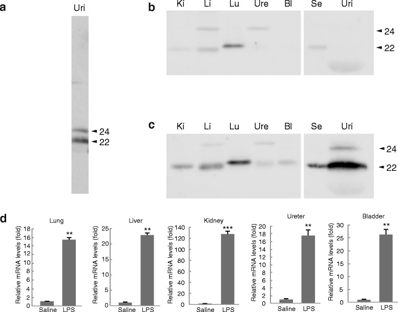
Fig. 1.

Detection of LCN2 in the urine from LPS-treated mice. Representative results of a Western blot analysis of the urinary LCN2 from LPS-treated mice (a). Male BALB/c mice were treated with (b) saline or (c) LPS (1 mg/kg i.p.) for 6 h. Tissue, serum, and urinary protein samples of 5 μg were subjected to Western blot analyses. Lu: lung, Li: liver, Ki: kidney, Ure: ureter, Bl: bladder, Se: serum, and Uri: urine. The numbers on the right sides of the figures indicate the molecular size of LCN2 (kDa). n = 6–8 for saline, and n = 8–10 for LPS treatment. (d) The LCN2 mRNA expression levels in peripheral tissues. The expression was normalized relative to that of β-actin, which served as an internal control. n = 5–7 for saline, and n = 4–5 for LPS treatment. Error bars represent the S.E.M. ** P < 0.01, and *** P < 0.001
