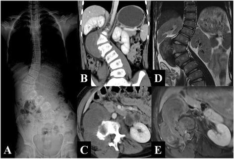
Fig. 1.

a The lower segment of the thoracic and lumbar spine existed a right tumefied thoracolumbar curve surrounding the first lumbar body with a Cobb angle of 33.7° and Ferguson angle of 69.4°. b, c Axial CT scans and coronary multi-plane reorganization demonstrated a right paravertebral irregular soft tissue mass with low and nonhomogeneous density, as well as uneven strength from the T12 to L2 vertebrae. The tumor grows through L1/2 right intervertebral foramen lesions to spinal canal. The intervertebral foramen becomes larger, and the right side of L2 vertebrae with irregular bone becomes corroded damage. d, e Axial enhanced MRI scan demonstrated the tumor is less homogeneous reinforcement, grows through L1/2 right intervertebral foramen lesions to spinal canal. The spinal shift to the left side due to cord compression
