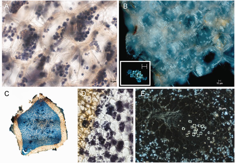
Fig. 3.—
Light microscopy of two different starch-containing tissues of Hydnora visseri. (A) Section of tepal, starch grains in cells stained with iodine–potassium iodide. (B) Section of tepal, showing starch grains under polarized light; inset: enlarged starch grains. (C) Transverse section of underground organ; stained with Astrablue-safranin; 5-merous organization of vascular system is visible. (D) Starch grains in the underground organ; stained with iodine-potassium iodide. (E) Starch grains and vascular bundle in underground organ under polarized light.

