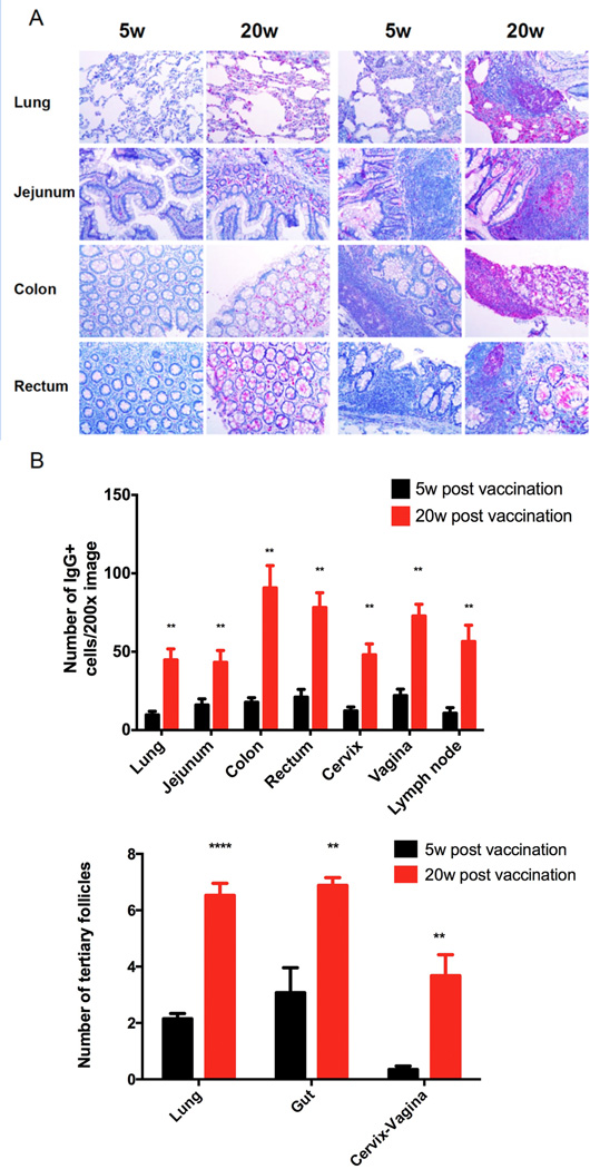
FIGURE 1.
General increase in IgG+ cells and ectopic follicles at the listed mucosal sites associated with the maturation of protection between 5 and 20 weeks (w). A. The left panel comparisons of 5 and 20w show increased numbers of red-magenta staining IgG+ plasma cells in the lung and gut between 5 and 20w; the right panels show the increases in ectopic follicles with IgG+ plasma cells. Image magnification 200×. B. Quantification of the increases at all mucosal sites. Significant P values (indicated by *) were determined by Student’s t test.

