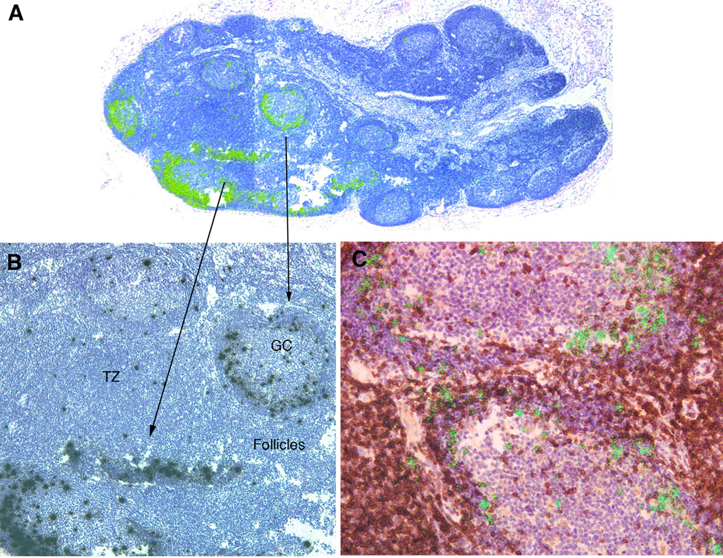
FIGURE 2.
SIVmac239Δnef replicates initially in TFH. A. Green viral RNA+ cells in epifluorescent reflected light images at the margins of lymphoid follicles 7 days post i.v. infection. B. Black viral+ cells in transmitted light. C. Double-labeled green viral RNA+ brown stained PD-1+ TFH cells at these locations. Viral RNA+ cells were also CD3+ and CD4+ (not shown).

