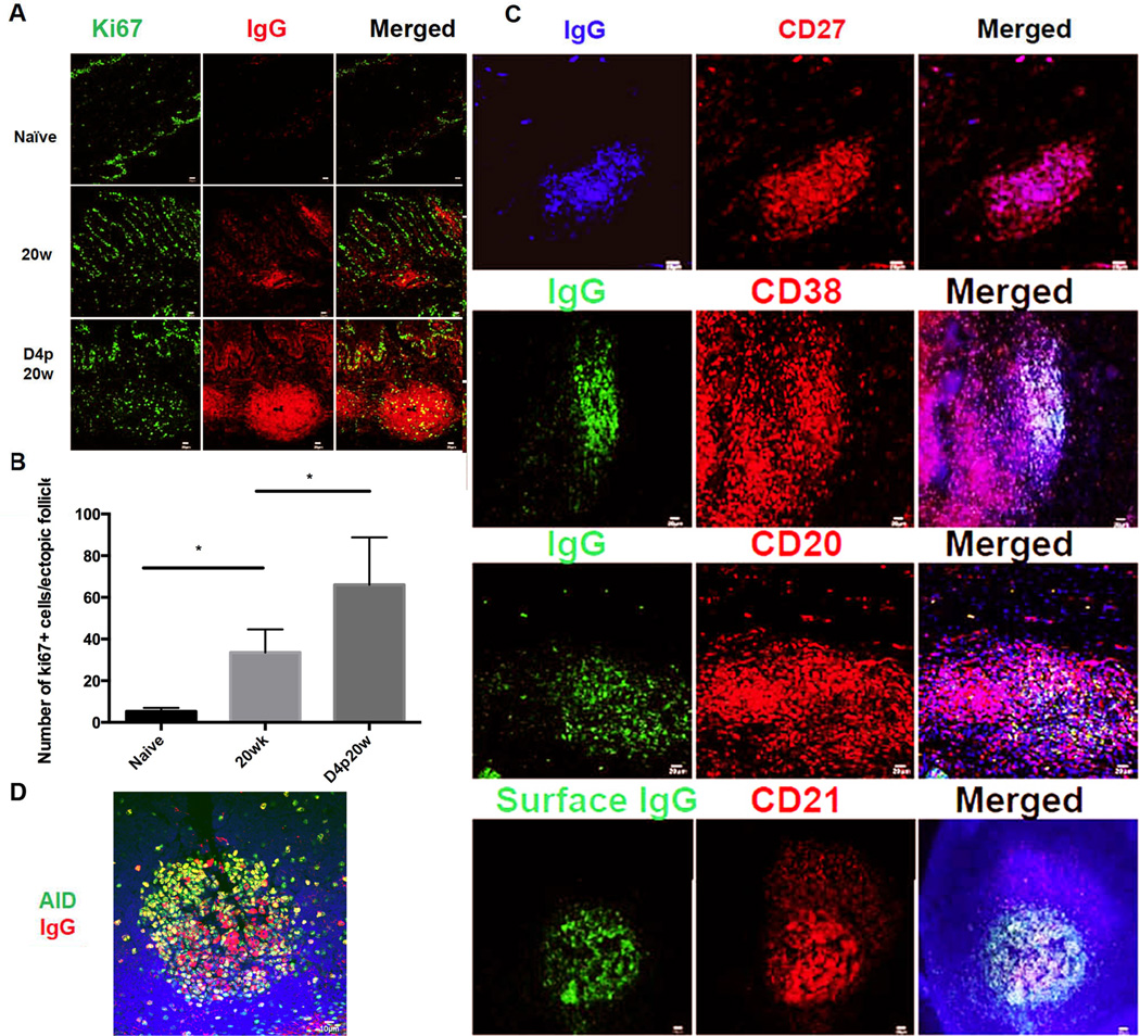
FIGURE 5.
Rapid induction of SIV-specific humoral immune response within vaginal ectopic follicles after WT SIVmac251 challenge. A. Increased red-stained IgG+ cells and follicles at 20w in vaccinated compared to unvaccinated animals. Most of the IgG+ cells are Ki67− (the green stained Ki67+ cells are primarily epithelial cells). Following vaginal challenge, Ki67+IgG+ cells increase beneath epithelium and particularly in the ectopic follicles. Representative images from n=4 for SIV-animals, n=4 for 20w and 3 for D4p20w animals. B. Quantification of increases in Ki67-cells at 2ow and D4p20w. Asterisks indicate significant increases. C. Phenotypic analysis of the IgG+ cells within ectopic follicles shows that they are CD38+CD27+CD20+ surface IgG+ plasmablasts. D. Fluorescent staining shows increased expression of activation-induced cytidine deaminase (AID) in the ectopic follicles after WT SIVmac251 challenge. Representative images were taken from animals at day 4 post-20 week vaccination.

