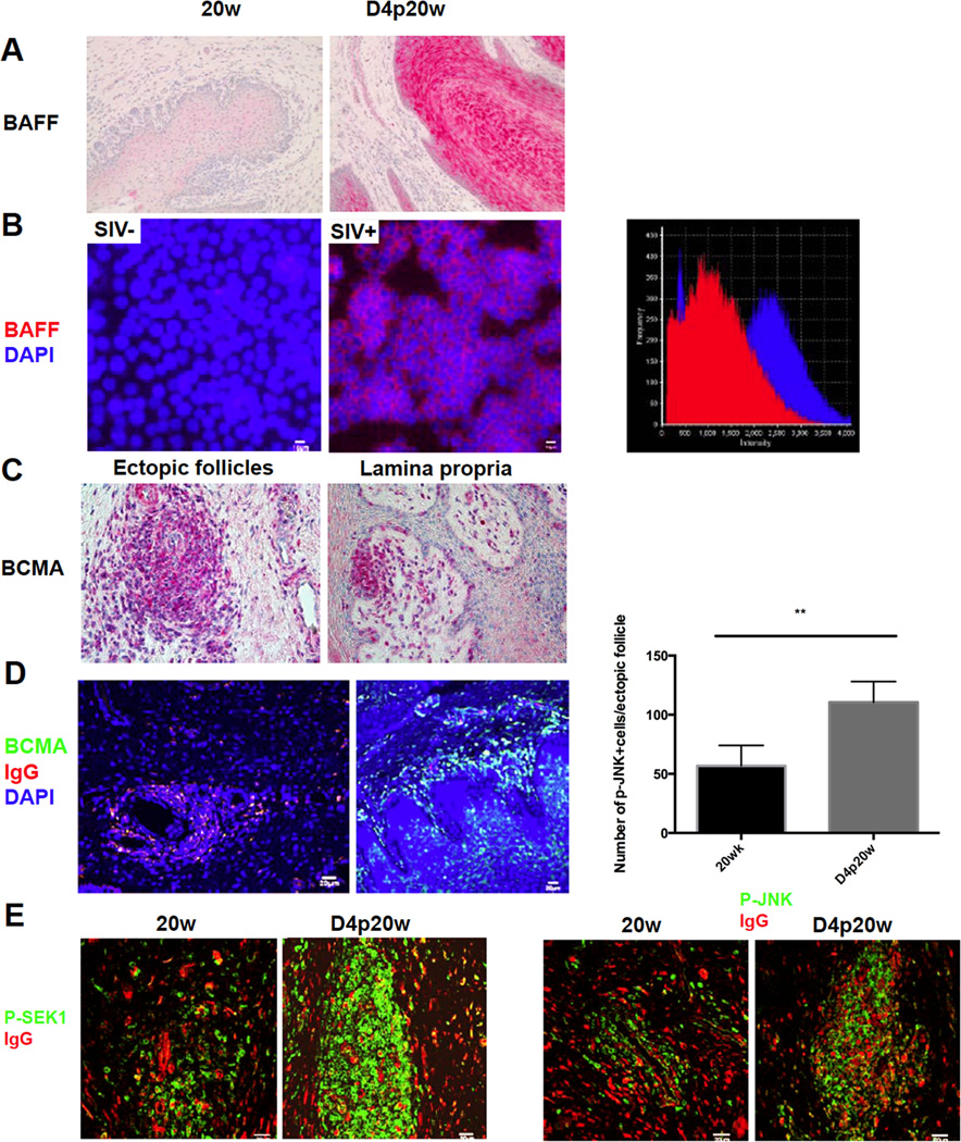
FIGURE 6.
Epithelial cells responses to promote the humoral immune response in the FRT. A, Increased BAFF expression in (red-stained) vaginal epithelium after WT SIVmac251 challenge. Image magnification: 200×. B. Fluorescent staining of BAFF (red) and DAPI (blue) shows that exposure to SIVmac251 for 2 days leads to up-regulation of BAFF in the HEC-1A epithelial cell line. Image on the far right shows the quantification by fluorescence intensity of BAFF staining in vehicle treated (SIV-) HEC-1A cells (red) and in SIV (SIV+) treated HEC-1A cells (blue). Result represents two independent experiments with duplicates of each treatment C, BCMA expression in magenta-stained cells within ectopic follicles and beneath the vaginal epithelium after WT SIVmac251 challenge. Representative images were taken from animals at day 5 and 7 post-20 week vaccination. Image magnification 400×. D, BMCA+ cells after challenge are IgG+ (yellow to yellow green). E. Fluorescent staining of phosphorylated-JNK and phosphorylated SEK1 (green) and IgG (red) shows that phosphorylated-JNK and phosphorylated SEK1, two key molecules in BAFF-BCMA signaling pathway, are both up-regulated after WT SIVmac251 challenge. Quantification in the panel above p-JNK staining shows the significant increases indicated by asterisks.

