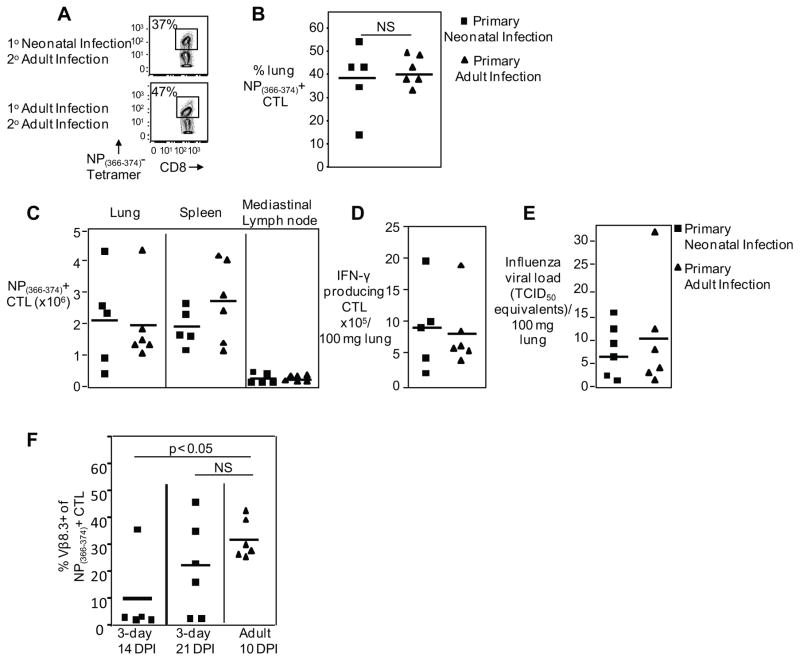
Figure 3. Mice infected during the neonatal period have a robust secondary CTL response.
Primary infection was performed in 3-day old neonatal or adult mice with PR8 influenza, 60 days later mice were challenged with H3N2 X31 recombinant strain and virus-specific CTLs were analyzed day 7 post-infection. Representative flow cytometry plots (A) and pooled data (B) showing percentage of pulmonary NP(366–374)-specific CTLs from mice either infected as neonates or adults and challenged as adults. (C) Pooled data showing NP(366–374)-specific CTL numbers in the lung, spleen and mediastinal lymph nodes from mice either infected as neonates or adults and challenged as adults. (D) IFN-γ producing CTL numbers shown in response to peptide stimulation after challenge. (E) Viral loads of neonates and adults after challenge were measured by real-time PCR and normalized per 100 mg of lung tissue. (F) Pooled data showing percentage of pulmonary NP(366–374)-specific CTLs which were Vβ 8.3 positive from mice either infected as neonates or adults and harvested at various time points. Symbols represent individual animals, horizontal line marking mean of each group (n= 5–6 mice per group, 2–3 independent experiments).

