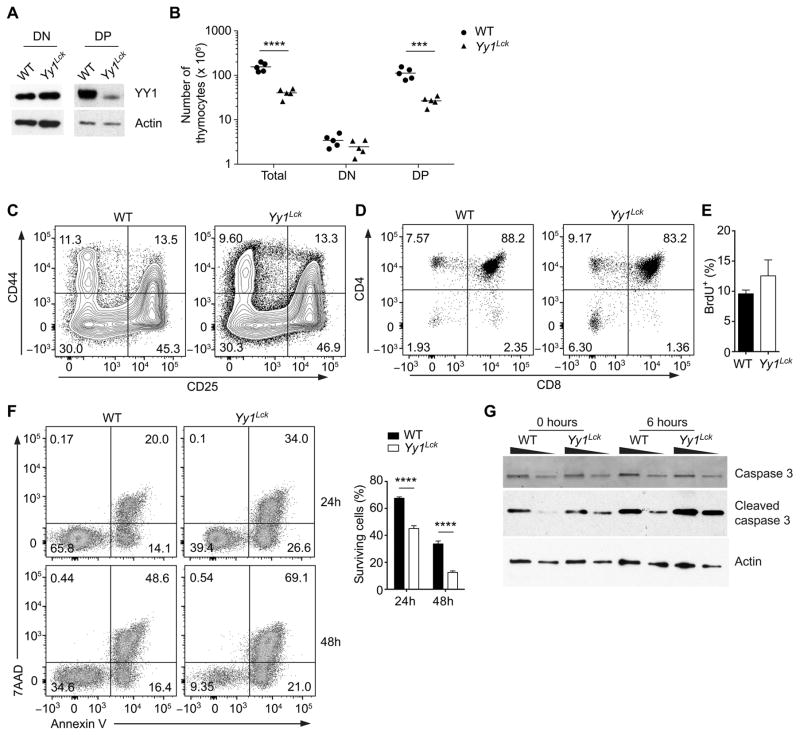
Figure 4. YY1 regulates the survival of DP thymocytes.
(A) Western blot of YY1 and actin in purified DN (CD4−CD8−Lin−) and DP thymocytes of Yy1f/f (WT) and Yy1f/f Lck-Cre (Yy1Lck) mice. Results are representative of two independent experiments. (B) Numbers of total, DN and DP thymocytes in WT and Yy1Lck mice. Each data point represents an individual mouse and the horizontal line indicates the mean. Statistical significance was evaluated by unpaired Student’s t-test with Holm-Sidak correction for multiple comparisons. (C) CD44 and CD25 staining of WT and Yy1Lck thymocytes pre-gated as CD4−CD8−Lin−. Results are representative of three independent experiments. (D) CD4 and CD8 staining of total thymocytes of WT and Yy1Lck mice. (E) Frequency of BrdU+ WT and Yy1Lck DP thymocytes following a 4 h pulse with BrdU. The mean ± SEM of three independent experiments is shown. (F) Sorted DP thymocytes were cultured in vitro for 24 or 48 h and stained with Annexin V and 7AAD (left). Mean ± SEM survival is presented for three WT and four Yy1Lck cultures (right). Statistical significance was evaluated by two-way ANOVA with Sidak’s multiple-comparison test. (G) Sorted DP thymocytes were analyzed for caspase 3 and cleaved caspase 3 by western blot either immediately ex vivo or after 6 h of in vitro culture. The wedges indicate 2-fold dilutions of cell extract. Data are representative of two independent experiments. ***P < 0.001, ****P < 0.0001.

